A Precise Diagnosis Can Make All the Difference
When a brain tumor is suspected, getting an accurate diagnosis is the first and most crucial step before beginning treatment. While MRI and CT scans can detect abnormalities in the brain, they cannot always determine the exact type of tumor. This is where Stereotactic Biopsy Brain Tumor plays a vital role.
Using advanced image-guided technology, a stereotactic brain biopsy allows neurosurgeons to obtain a small tissue sample from the tumor with exceptional precision. This minimally invasive procedure helps doctors identify the nature of the tumor and develop the most effective treatment plan while minimizing risks to healthy brain tissue.
Dr. Manish Baldia is a leading neurosurgeon in Mumbai with expertise in stereotactic procedures, brain tumor surgery, functional neurosurgery, and minimally invasive brain and spine surgeries. His focus on precision, patient safety, and advanced technology has helped numerous patients receive timely and accurate brain tumor diagnoses.
Why Is an Accurate Brain Tumor Diagnosis So Important?
Every brain tumor is different. Some tumors are benign, while others may be cancerous or rapidly growing. Even tumors that appear similar on scans can require completely different treatments.
An accurate diagnosis helps doctors:
- Identify the exact type of brain tumor
- Determine whether the tumor is benign or malignant
- Understand how aggressive the tumor is
- Plan surgery more effectively
- Decide whether radiation or chemotherapy is required
- Avoid unnecessary procedures
Without a tissue diagnosis, choosing the right treatment becomes significantly more challenging.
What Is a Stereotactic Brain Biopsy?
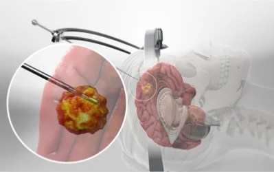
A Stereotactic Brain Biopsy is a minimally invasive neurosurgical procedure that uses computer-assisted navigation together with MRI or CT imaging to accurately reach a brain lesion.
Instead of performing a large open surgery, the neurosurgeon makes a very small opening in the skull and inserts a specialized biopsy needle to collect tiny tissue samples from the affected area.
These samples are then examined under a microscope to confirm:
- Tumor type
- Tumor grade
- Whether the growth is cancerous
- Molecular characteristics
- Future treatment options
This approach provides highly accurate results with minimal disruption to surrounding brain tissue.
When Do Doctors Recommend a Stereotactic Biopsy?
Not every brain tumor can be removed immediately. Some tumors develop deep inside the brain or are located near areas that control speech, movement, vision, or memory.
A stereotactic biopsy is commonly recommended when:
- The tumor is difficult to access safely.
- Surgery carries a high risk.
- The diagnosis remains uncertain after MRI or CT scans.
- The lesion may represent an infection rather than a tumor.
- Treatment decisions depend on laboratory confirmation.
For many patients, this procedure serves as the safest way to establish a clear diagnosis before beginning treatment.
Common Symptoms That May Require Further Evaluation
Although symptoms vary depending on the location of the tumor, some of the most common warning signs include:
- Persistent headaches
- Frequent seizures
- Weakness in the arms or legs
- Difficulty speaking
- Vision problems
- Memory loss
- Loss of balance
- Personality changes
- Nausea or vomiting
If these symptoms continue or worsen, consulting an experienced neurosurgeon is essential.
How Is the Procedure Performed?
One of the biggest advantages of stereotactic biopsy is its precision.
Step 1 – Advanced Brain Imaging
MRI or CT scans are performed to identify the exact location of the abnormal tissue.
Step 2 – Computer-Guided Planning
Specialized stereotactic software calculates the safest and most accurate pathway to the lesion.
Step 3 – Tissue Collection
A tiny incision is made, and a biopsy needle is carefully guided to the tumor to collect tissue samples.
Step 4 – Laboratory Examination
Expert pathologists analyze the sample to determine the exact diagnosis, helping doctors plan the most appropriate treatment.
The procedure usually requires only a small incision and often allows for a quicker recovery compared to open brain surgery.
Benefits of Stereotactic Brain Biopsy
Modern stereotactic techniques offer several advantages over traditional diagnostic surgery.
Patients benefit from:
- Highly accurate diagnosis
- Minimally invasive approach
- Small incision
- Less pain
- Reduced bleeding
- Lower complication rates
- Shorter hospital stay
- Faster recovery
- Excellent precision for deep-seated tumors
These benefits make stereotactic biopsy one of the preferred diagnostic procedures for complex brain lesions.
What Happens After the Biopsy?
Once the laboratory report confirms the diagnosis, Dr. Manish Baldia develops an individualized treatment plan.
Depending on the findings, treatments may include:
- Brain Tumor Surgery
- Awake Brain Tumor Surgery
- Radiation Therapy
- Chemotherapy
- Targeted Therapy
- Gamma Knife Radiosurgery
- CyberKnife Treatment
- Regular observation for non-aggressive tumors
Every treatment plan is carefully tailored according to the patient’s condition and long-term health goals.
Meet Dr. Manish Baldia – Advanced Neurosurgical Care You Can Trust
Choosing the right neurosurgeon is one of the most important decisions for patients diagnosed with a brain tumor.
Dr. Manish Baldia is a highly experienced neurosurgeon specializing in:
- Stereotactic Brain Biopsy
- Brain Tumor Surgery
- Awake Brain Tumor Surgery
- Deep Brain Stimulation (DBS)
- Functional Neurosurgery
- Stereotactic Procedures
- Spine Surgery
- Movement Disorder Surgery
After completing his MCh in Neurosurgery from the prestigious Christian Medical College (CMC), Vellore, he underwent advanced fellowship training in Functional Neurosurgery in London, enabling him to perform some of the most complex neurological procedures with precision and confidence.
His patient-centered approach ensures that every individual receives comprehensive evaluation, advanced treatment, and compassionate care throughout their journey.
Advanced Facilities for Comprehensive Brain Tumor Care
Patients treated by Dr. Manish Baldia have access to modern neurosurgical facilities equipped with advanced technology, including:
- High-resolution MRI & CT Imaging
- Computer-Assisted Stereotactic Navigation
- Modern Neurosurgical Operating Rooms
- Experienced Neuro-Anesthesia Team
- Intensive Care Support
- Advanced Neuropathology Services
- Postoperative Rehabilitation
- Multidisciplinary Neurological Care
These advanced resources contribute to accurate diagnosis, safer procedures, and improved recovery outcomes.
Take the First Step Towards an Accurate Diagnosis
A brain tumor diagnosis should never be based on assumptions. With Stereotactic Biopsy Brain Tumor in Mumbai, patients can receive a precise diagnosis through a minimally invasive procedure that guides the most effective treatment plan.
If you or someone you love has been advised to undergo a brain biopsy, consult Dr. Manish Baldia for expert evaluation and advanced neurosurgical care.
Dr. Manish Baldia – Expert Neurosurgeon | Specialized in DBS, Spine Surgery & Stereotactic Procedures
Address:
Lalbaug Diagnostics, Dr. Babasaheb Ambedkar Marg, near Ladu Samrat Vada Pav, opposite Avighna IX, Lal Baug, Parel, Mumbai – 400012
Phone: 9676767507