
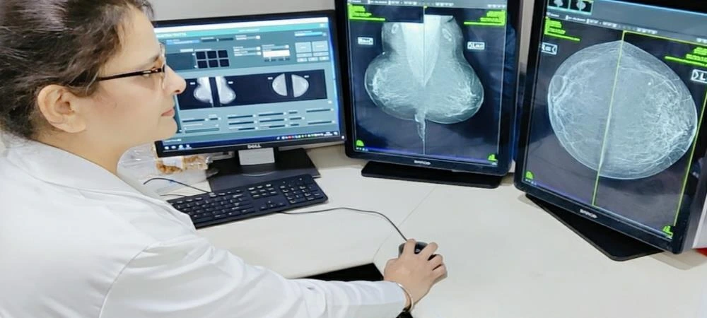
During my review of breast health services in Delhi, I found that discovering a breast lump or an unusual breast change can understandably cause anxiety. However, not every breast lump is cancerous, and a structured diagnostic approach can help determine the cause. Triple Assessment of Breast in Delhi is a comprehensive evaluation that combines clinical examination, breast imaging and tissue diagnosis when required. Dr. R K Saggu, an oncoplastic breast surgeon associated with Max Super Speciality Hospital, focuses on breast cancer and non-cancerous breast conditions, with extensive experience in breast diagnosis and surgery.
The purpose of triple assessment is to evaluate a breast concern systematically rather than relying on a single test. Depending on the findings, patients may undergo clinical examination, ultrasound or mammography, followed by a needle biopsy if an abnormality is identified.
Triple Assessment of Breast in Delhi: Three Steps of Evaluation
Triple Assessment of Breast in Delhi brings together three important components: clinical assessment, breast imaging and pathological assessment. Using these methods together can provide a more complete understanding of a breast lump or other breast abnormality.
1. Clinical Breast Examination
The first part involves a detailed medical history and physical examination of the breasts and armpits.
The doctor may ask about:
- When the lump or breast change was first noticed
- Whether the lump is painful or painless
- Previous breast problems
- Family history of breast cancer
- Menstrual and reproductive history
- Previous breast imaging or biopsies
- Other relevant health conditions
Dr. R K Saggu’s breast diagnosis approach includes assessing risk factors and examining both breasts and the armpit region.
A clinical examination can help determine whether further investigation is required and which imaging approach may be appropriate.
2. Breast Imaging
The second component is breast imaging. Depending on the patient’s age, symptoms and clinical findings, this may involve ultrasound, mammography or both.
Ultrasound can be particularly useful for assessing breast lumps and determining whether a lesion appears solid or fluid-filled. Mammography is an important imaging tool, and the information on Dr. R K Saggu’s website notes that women over 40 may require mammography as part of assessment.
Additional imaging, such as breast MRI, may be recommended in selected cases when more information is needed.
3. Needle Biopsy and Pathological Assessment
If clinical examination or imaging identifies a suspicious abnormality, a tissue sample may be recommended.
A breast biopsy involves removing cells or tissue from the area of concern so that a pathologist can examine it under a microscope. According to Dr. R K Saggu’s diagnostic information, biopsy is the definitive method for determining whether cells are cancerous.
Different biopsy techniques may be used depending on the nature and location of the lesion.
Types of Breast Biopsy
Several biopsy methods are available, and the appropriate technique depends on the individual case.
Fine Needle Aspiration
Fine needle aspiration, or FNA, uses a thin needle to obtain fluid and cells from a breast lump. It is a relatively simple procedure, but additional biopsy may be necessary in some cases because FNA may not provide enough information to determine whether a cancer is invasive or non-invasive.
Core Needle Biopsy
A core biopsy uses a hollow needle to remove small cylindrical samples of breast tissue. It is generally performed as an outpatient procedure under local anaesthesia.
The tissue is sent to a pathologist for examination. A core biopsy can help confirm cancer and identify its type. Dr. R K Saggu’s website states that ER, PR and HER2neu immunohistochemistry markers are routinely assessed for breast cancer patients to help guide subsequent treatment planning.
Image-Guided Biopsy
When an abnormality is visible on imaging but cannot be easily felt, image guidance can help target the biopsy accurately. Depending on the imaging modality, a biopsy may be ultrasound-guided, stereotactic or MRI-guided.
Vacuum-Assisted Breast Biopsy
Vacuum-assisted breast biopsy uses an image-guided probe to obtain multiple tissue samples. It can remove more tissue than a conventional core needle biopsy and may be used for selected breast abnormalities.
What Happens After the Biopsy?
A biopsy sample is examined by a pathologist, and the report helps determine the nature of the breast abnormality.
Dr. R K Saggu’s website states that biopsy reports are reviewed with the patient and family and explained in detail. When cancer is diagnosed, treatment planning is undertaken according to the patient’s individual circumstances. Her website also describes discussion of cases in a multidisciplinary meeting before developing a personalised treatment plan.
This step is important because breast cancer is not one single disease. Different breast cancers can have different biological characteristics, stages and treatment requirements.
Understanding Breast Cancer Staging
If breast cancer is confirmed, additional investigations may be recommended to determine whether and how far the disease has spread.
Possible staging investigations include:
- Breast MRI
- CT scan
- PET scan
- Bone scan
- Chest X-ray
- Abdominal ultrasound
Not every patient requires every test. The appropriate investigations depend on the clinical and pathological findings.
Staging helps doctors understand the extent of disease and develop an appropriate treatment strategy.
When Should You Seek Breast Assessment?
Women should seek medical evaluation when they notice a new or persistent breast change. This may include:
- A new breast lump
- Changes in breast shape or size
- Skin changes
- Nipple changes
- Nipple discharge
- A lump in the armpit
- Persistent breast discomfort
- Changes noticed during breast self-awareness checks
A breast lump does not automatically indicate cancer. Benign conditions such as fibroadenomas and other non-cancerous breast diseases can also produce lumps.
The important step is to have an appropriate clinical assessment rather than attempting to determine the cause based on symptoms alone.
Dr. R K Saggu: Qualifications and Clinical Experience
Dr. Rajinder Kaur Saggu is an oncoplastic breast surgeon with 18 years of overall surgical experience and 13 years of professional experience as a surgical oncologist, according to her professional profile. Her practice focuses particularly on breast cancer and non-cancerous breast conditions, and she has experience performing different types of breast surgeries.
Her qualifications include MBBS from Government Medical College, Miraj, MS in Surgery from Government Medical College, Patiala, and a Breast Fellowship from Tata Memorial Hospital, Mumbai. She also completed advanced breast oncoplasty training at the University of East Anglia in the UK and breast reconstruction training at MD Anderson Cancer Center in Houston, Texas.
Her training additionally includes endoscopic and robotic breast surgery through workshops in Paris and Abu Dhabi. Her professional profile notes experience with endoscopic breast surgery in selected cases and expertise in vacuum-assisted breast biopsy and excision for fibroadenomas and other benign breast conditions.
Clinic Environment and Breast Care Approach
Dr. R K Saggu’s clinic is listed at 37/3, East Patel Nagar, New Delhi, while her hospital affiliation is listed as Max Super Speciality Hospital, Vaishali, Ghaziabad. Her practice includes breast cancer diagnosis, benign breast disease, breast cancer surgery and post-surgery care.
Her diagnostic approach emphasises helping patients understand each stage of assessment. The website also notes that patients are welcome to attend appointments with a partner, friend or relative, which can be helpful when discussing investigations or possible diagnoses.
Beyond clinical care, Dr. R K Saggu is involved in breast cancer awareness initiatives. She and her husband established Guru Nanak Cancer Care Trust in 2012 to provide services and support to underprivileged women while promoting awareness about early breast cancer detection.
Expertise in Breast Cancer and Oncoplastic Surgery
Dr. R K Saggu’s professional experience spans breast oncology, reconstructive breast surgery and oncoplastic techniques. Her career includes work at Lady Hardinge Medical College, Tata Memorial Hospital, MD Anderson Cancer Center and Indraprastha Apollo Hospitals. She has also been associated with Max Institute of Cancer Care, Vaishali, since 2023.
For patients searching for the Best Breast Cancer Surgeon in Delhi, relevant factors include specialist training, breast-specific experience, diagnostic expertise, surgical capabilities and the ability to develop an individual treatment plan.
Patients looking for a Breast Cancer Surgeon in Delhi should also understand that diagnosis and treatment are separate stages. A suspicious breast lump does not necessarily mean cancer, and appropriate imaging and pathology are essential before treatment decisions are made.
Benefits and Considerations of Triple Assessment
The major benefit of triple assessment is that it combines information from clinical examination, imaging and pathology rather than relying on one diagnostic method.
This approach can:
- Evaluate a newly detected breast lump
- Identify suspicious abnormalities
- Help distinguish benign from malignant conditions
- Guide the need for biopsy
- Support treatment planning when cancer is confirmed
However, individual patients may require different tests. Not every person with a breast lump will require all available imaging or biopsy techniques. The diagnostic plan should be based on clinical findings and specialist assessment.
Consult Dr. R K Saggu
If you have noticed a breast lump or any unusual breast change, consider scheduling a consultation with Dr. R K Saggu for an appropriate breast assessment.
Get Directions to the Clinic – Max Super Speciality Hospital
Early evaluation does not mean that a lump is cancerous. It provides an opportunity to understand the cause through appropriate examination, imaging and pathology when required.
Frequently Asked Questions
1. What is Triple Assessment of the Breast?
Triple assessment combines clinical breast examination, breast imaging such as ultrasound or mammography, and tissue diagnosis through biopsy when indicated. It provides a structured approach to evaluating breast lumps and other abnormalities.
2. Does every breast lump require a biopsy?
No. The need for biopsy depends on the clinical examination and imaging findings. If an abnormality appears suspicious, a needle biopsy may be recommended to establish a definitive diagnosis.
3. Is a breast biopsy painful?
Many needle biopsies are performed using local anaesthesia. Patients may experience some pressure or brief discomfort, but the experience varies between individuals. Dr. R K Saggu’s website describes core needle biopsy as an outpatient procedure performed under local anaesthesia.
4. What happens if the biopsy confirms breast cancer?
If cancer is confirmed, additional assessment may be performed to determine its stage and biological characteristics. Treatment can then be planned according to the individual’s diagnosis, stage and overall health. Dr. R K Saggu’s website describes multidisciplinary discussion and personalised treatment planning.